Scanning Electron Microscope
Focused Ion Beam Scanning Electron Microscope DB500
Description
Features
Specifications
Description
CIQTEK DB500 is a Field Emission Scanning Electron Microscope with a Focused Ion Beam column for nano analysis and specimen preparation, which is applied with “SuperTunnel” technology, low aberration, and magnetic-free objective lens design, with low-voltage and high-resolution ability that ensures its nano-scale analytical capability. The ion column facilitates a Ga+ liquid metal ion source with a highly stable and high-quality ion beam to ensure nano-fabrication capability.
DB500 is equipped with an integrated nano-manipulator, gas injection system, electrical anti-contamination mechanism for the objective lens, and 24 expansion ports, making it an all-around nano-analysis and fabrication platform with comprehensive configurations and expandability.
Features
SuperTunnel” Electron Optics technology with a magnetic-free objective lens, suitable for high-resolution imaging, and compatible with magnetic specimen imaging.
The focused ion beam column which outputs a highly stable, high-quality ion beam, suitable for high-quality nano-fabrication and TEM specimen preparation.
A piezoelectrically-driven manipulator located in the specimen chamber with an integrated control system for precise handling.
Independently developed system with strong expandability. The integrated ion source assembly design for quick ion source exchange. Excellent service, supported by an included three-year warranty.
Specifications
| Electron Beam System | Electron Gun Type | High Brightness Schottky Field Emission Electron Gun |
| Resolution | 1.2 nm@15 kV | |
| Acceleration Voltage | 20 V~30 kV | |
| Ion Beam System | Ion Source Type | Liquid Gallium Ion Source |
| Resolution | 3 nm@30 kV | |
| Acceleration Voltage | 500 V ~ 30 kV | |
| Specimen Chamber | Vacuum System | Fully Automatic Control, Oil-free Vacuum System |
| Cameras | Three Cameras (Optical navigation + chamber monitor x2) | |
| Stage Type | 5-axis Mechanical Eucentric Specimen Stage | |
| Stage Range | X=110 mm, Y=110 mm, Z=65 mm T: -10°~+70°, R:360° | |
| Detectors and Extensions | Standard | In-lens Detector Everhart-Thornley Detector (ETD) |
| Optional | Retractable Back-Scattered Electron Detector (BSED) Retractable Scanning Transmission Electron Microscopy Detector (STEM) Energy Dispersive Spectrometer (EDS) Electron Backscatter Diffraction Pattern (EBSD) Nano-manipulator Gas Injection System Plasma Cleaner Specimen Exchange Loadlock Trackball & Knob Control Panel | |
| Software | Languages | English |
| Operating System | Windows | |
| Navigation | Nav-Cam, Gesture Quick Navigation | |
| Automatic Functions | Auto Brightness & Contrast, Auto Focus, Auto Stigmator |
Ultra-high Resolution Field Emission Scanning Electron Microscope | SEM5000X
Description
Features
Specifications
Description
CIQTEK SEM5000X is an ultra-high resolution Field Emission Scanning Electron Microscope (FE-SEM) with breakthrough resolution of 0.6 nm@15 kV and 1.0 nm@1 kV.
Benefiting from the upgraded column engineering process, “SuperTunnel” technology, and high-resolution objective lens design, SEM5000X can achieve further improvements in low-voltage imaging resolution. The specimen chamber ports extend to a number of 16, and the specimen exchange load-lock supports up to 8-inch wafer size (maximum diameter 208 mm), greatly expanding applications’ coverage. The advanced scanning modes and enhanced automated functions bring stronger performance and an even more optimized experience.
Features
Ultra-high Resolution
(0.6 nm@15 kV).
Mechanical Eucentric Specimen Stage.
Specimen Exchange Loadlock (Maximum 8″).
Specifications
| Key Parameters | Resolution | 0.6 nm @ 15 kV, SE 1.0 nm @ 1 kV, SE |
| Acceleration Voltage | 20 V ~ 30 kV | |
| Magnification | 1 ~ 2,500,000 x | |
| Electron Gun Type | Schottky Field Emission Electron Gun | |
| Specimen Chamber | Vacuum System | Fully Automated Control |
| Cameras | Dual Cameras (optical navigation + chamber monitor) | |
| Stage Type | 5-Axis Mechanical Eucentric Specimen Stage | |
| Stage Range | X=110 mm, Y=110 mm, Z=65 mm T: -10*~+70°, R: 360° | |
| Detectors and Extensions | Standard | In-lens Detector Everhart-Thornley Detector (ETD) |
| Optional | Retractable Back-Scattered Electron Detector(BSED) Retractable Scanning Transmission Electron Microscopy Detector (STEM) Energy Dispersive Spectrometer (EDS) Electron Backscatter Diffraction Pattern (EBSD) Specimen Exchange Loadlock (4″ & 8″ optional) Trackball & Knob Control Panel Specimen Stage Tandem Deceleration Magnetic Field & Acoustic Noise Enclosure System(SEMI Certified) | |
| Software | Languages | English |
| Operating System | Windows | |
| Navigation | Nav-Cam, Gesture Quick Navigation | |
| Automatic Functions | Auto Brightness & Contrast, Auto Focus, Auto Stigmator |
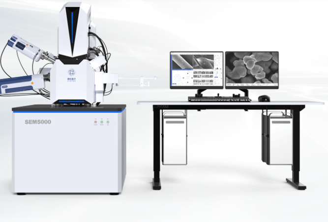
Field Emission Scanning Electron Microscope | SEM5000
Description
Features
Specifications
Description
CIQTEK SEM3200 is a high-performance tungsten filament scanning electron microscope. It has excellent image quality, low vacuum mode compatibility, high resolution images in different fields of view. The depth of field is large and the image is rich in stereo.CIQTEK SEM5000 is a field emission scanning electron microscope with high-resolution imaging and analysis ability, supported by abundant functions, benefits from advanced electron optics column design, with high-pressure electron beam tunnel technology (SuperTunnel), low aberration, and non-immersion objective lens, achieves low voltage high-resolution imaging, the magnetic specimen can also be analyzed.
With optical navigation, automated functionalities, carefully designed human-computer interaction user interface, and optimized operation and use process, no matter if you are an expert or not, you can quickly get started and complete high-resolution imaging and analysis work.
Features
• High-resolution imaging at low accelerating voltage.
• Electromagnetic composite objective improves low-voltage resolution and enables magnetic specimen observation.
• High-pressure tunneling technology (SuperTunnel) ensures low voltage resolution.
• The electronic optical path without crossover effectively reduces system aberration and improves resolution.
• Water-cooled constant temperature objective lens, to ensure the stability, reliability, and repeatability of the objective lens work.
• Magnetic deflected six-hole various aperture system, with automatic switchable apertures, no mechanical adjustment needed, achieves high-resolution imaging or large beam analysis mode through a click-away fast switching.
Specifications
| Key Parameters | Resolution | 0.9 nm @ 15 kV, SE 1.3 nm @ 1.0 kV, SE 0.8 nm @ 30 kV, STEM |
| Acceleration Voltage | 20 V ~ 30 kV | |
| Magnification | 1 ~ 2,500,000 x | |
| Electron Gun Type | High brightness Schottky Field Emission Electron Gun | |
| Specimen Chamber | Vacuum System | Fully Automatic Control, Oil-free Vacuum System |
| Camera | Dual Cameras (Optical navigation + chamber monitor) | |
| Stage Range | X: 125 mm, Y: 125 mm, Z: 50 mm T: -10°~ +90°, R: 360° (*Optional extra-large chamber version available) | |
| Detectors and Extensions | Standard | Inlens SE Detector Everhart-Thornley Detector (ETD) |
| Optional | Flat Insertion Medium Angle Backscattered Electron Detector STEM Automatic Retractable Scanning Transmission Electron Detector Specimen Exchange Loadlock Fast Beam Blanker Energy Dispersive Spectroscopy (EDS/EDX) Electron Backscatter Diffraction Pattern (EBSD) Electron-beam-induced Current (EBIC) Cathodoluminescence (CL) In situ Tensile Stage Nano Manipulator Large-scale Image Stitching Trackball & Knob Control Panel | |
| Software | Language | English |
| Operating System | Windows | |
| Navigation | Nav-Cam, Gesture Navigation | |
| Automatic Functions | Auto Brightness & Contrast, Auto Focus, Auto Stigmator |
Field Emission Scanning Electron Microscope SEM4000
Description
Features
Specifications
Description
CIQTEK SEM4000 is an analytical field emission scanning electron microscope equipped with a high-brightness long-life Schottky field emission electron gun.
With the three-stage condenser electron optics column design and the large continuously adjustable beam current, SEM4000 delivers advantages in EDS, EBSD, WDS, and other analytical applications. The system supports low vacuum mode, which can help directly observe poorly conductive or even non-conductive specimens. Standard optical navigation mode, as well as an intuitive user operation interface, makes your analysis work easy.
Features
Expandability: SEM SE\BSE\EDS\EDX\EBSD, etc.
Optical Navigation: Quickly locates targeted Region of Interest (ROI).
Large FOV Mapping: Fully automatic image stitching to map a large field of view.
Mixing Image (SE+BSE): Observe the specimen compositional and surface topographic information in one image.
Dual Anode (Tetrode): Dual anode emission system design provides excellent resolution under low landing energy.
Low Vacuum Mode: Provide specimen surface morphology information in low vacuum, switchable vacuum state with one click.
Specifications
| |||||||||||||||||||||||||||||||||||||||||||||||||||||||||||||||||||||||||||||||||||||||||||||||||||||||||||||||||||||
Tungsten Filament Scanning Electron Microscope | SEM3200
Description
Features
Specifications
Description
CIQTEK SEM3200 is a high-performance tungsten filament scanning electron microscope. It has excellent imaging quality capabilities in both high and low vacuum modes. It also has a large depth of field with a user-friendly interface to enable users to characterize specimens and explore the world of microscopic imaging and analysis.
Features
Equipped with high brightness and long-life Schottky field emission electron gun
• High resolution, better than 1nm resolution at 30 kV
• Three-stage condenser lens design, wide beam current adjustable range
• *High-performance low vacuum secondary electron detectors, observe poorly conductive or non-conductive specimens
• Non-immersion magnetic field free objective lens design, can directly observe magnetic specimens
• Standard optical navigation mode
Specifications
| Models | SEM3200A | SEM3200 | ||
| Electro-Optical Systems | Electron Gun | Pre-aligned Medium-sized Hairpin-type Tungsten Filament | ||
| Resolution | High Vaccum | 3 nm @ 30 kV (SE) | ||
| 4 nm @ 30 kV (BSE) | ||||
| 8 nm @ 3 kV (SE) | ||||
| *Low Vaccum | 3 nm @ 30 kV (SE) | |||
| Magnification | 1-300,000x (Film Magnification) | |||
| 1-1000,000x (Screen Magnification) | ||||
| Acceleration Voltage | 0.2 kV ~ 30 kV | |||
| Probe Current | ≥1.2μA, Real-time display | |||
| Imaging Systems | Detector | Everhart-Thornley Detector (ETD) | ||
| *Backscattered electron detector (BSED), *Low vacuum secondary electron detector, *Energy spectrometer EDS, etc. | ||||
| Image Format | TIFF, JPG, BMP, PNG | |||
| Vacuum System | Vacuum Model | High Vacuum | Better than 5×10⁻⁴ Pa | |
| Low Vacuum | 5 ~ 1000 Pa | |||
| Control Mode | Fully Automated Control | |||
| Specimen Chamber | Camera | Optical Navigation | ||
| Monitoring in the Specimen Chamber | ||||
| Specimen Table | Three Axis Automatic | Five Axis Automatic | ||
| Stage Range | X: 120 mm | X: 120 mm | ||
| Y: 115 mm | Y: 115 mm | |||
| Z: 50 mm | Z: 50 mm | |||
| / | R: 360° | |||
| / | T: -10° ~ +90° | |||
| Software | Operating System | Windows | ||
| Navigations | Optical Navigation, Gesture Quick Navigation | |||
| Automatic Functions | Auto Brightness & Contrast, Auto Focus, Automatic Stigmator | |||
| Special Functions | Intelligence Assisted Image Astigmatism Correction *Large-FOV Image Stitching (Optional) | |||
| Installation Requirements | Space | L≥ 3000 mm, W ≥ 4000 mm,H ≥ 2300 mm | ||
| Temperature | 20°C (68°F) ~ 25°C (77°F) | |||
| Humidity | ≤ 50 % | |||
| Power Supply | AC 220 V(±10 %), 50 Hz, 2 kVA | |||